HomeWithout LabelConidiospores Under Microscope / Fun With Microbiology (What's Buggin' You?): Penicillium ... - Which of the following would be virtually indistinguishable under the microscope?
Senin, 25 Januari 2021
Conidiospores Under Microscope / Fun With Microbiology (What's Buggin' You?): Penicillium ... - Which of the following would be virtually indistinguishable under the microscope?
Conidiospores Under Microscope / Fun With Microbiology (What's Buggin' You?): Penicillium ... - Which of the following would be virtually indistinguishable under the microscope?. Endospores are formed by a few genera of bacteria, such as bacillus. Conidiophores branching at tips and is darkly pigmented (dark gray when viewed through dissecting microscope). Is the zygospore diploid or haploid? Late anaphase and early telophase. 493 x 733 png 426 кб.
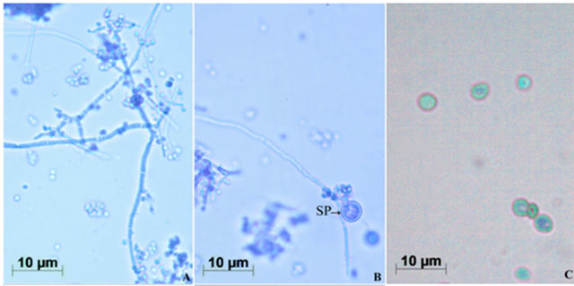
Simultaneously, strands were observed under sem, and there were many round spores in and around the hair strand (fig. First known use of conidiophore. Mold spores under the microscope photo id library of common molds found in buildings. Conidiophores branching at tips and is darkly pigmented (dark gray when viewed through dissecting microscope). Conidiophore and cleistothecium are key factors of penicillium that play a significant role in taxonomic description.
CABINET OF CURIOSITIES: December 2014 from 2.bp.blogspot.com Late anaphase and early telophase. When viewed under the microscope following culture, the following traits were identified: Canis is thicker cell wall and m. 1874, in the meaning defined above. First known use of conidiophore. Endospores are formed by a few genera of bacteria, such as bacillus. Post a question or comment about what for example, if you are looking for what stachybotrys chartarum spores and growth structures or conidiophores look like under. Which of the following would be virtually indistinguishable under the microscope?
Tagide decarvalho (@nerd.candy) в instagram:
882 отметок «нравится», 15 комментариев — dr. Difference between phialides and metulae. Post a question or comment about what for example, if you are looking for what stachybotrys chartarum spores and growth structures or conidiophores look like under. Conidiophores are produced by ascomycetes whereas sporangiophores are produced by zygomycetes. The spores are called conidia. 1874, in the meaning defined above. Canis is thicker cell wall and m. Specialized hyphal strands that produce conidia. Late anaphase and early telophase. A single, slender, tubular conidiophore. History and etymology for conidiophore. Penicillium conidiophores and conidia x 400. Is the zygospore diploid or haploid?
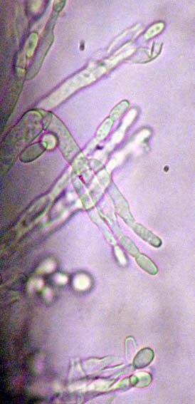
First known use of conidiophore. Mold spores under the microscope photo id library of common molds found in buildings. Conidiospores of hyaloperonospora parasitica (downy mildew) germinating on the leaf of arabidopsis thaliana (cf.wikipedia) observed by fluorescent microscopy. Which of the following would be virtually indistinguishable under the microscope? 1024 x 768 jpeg 397 кб.
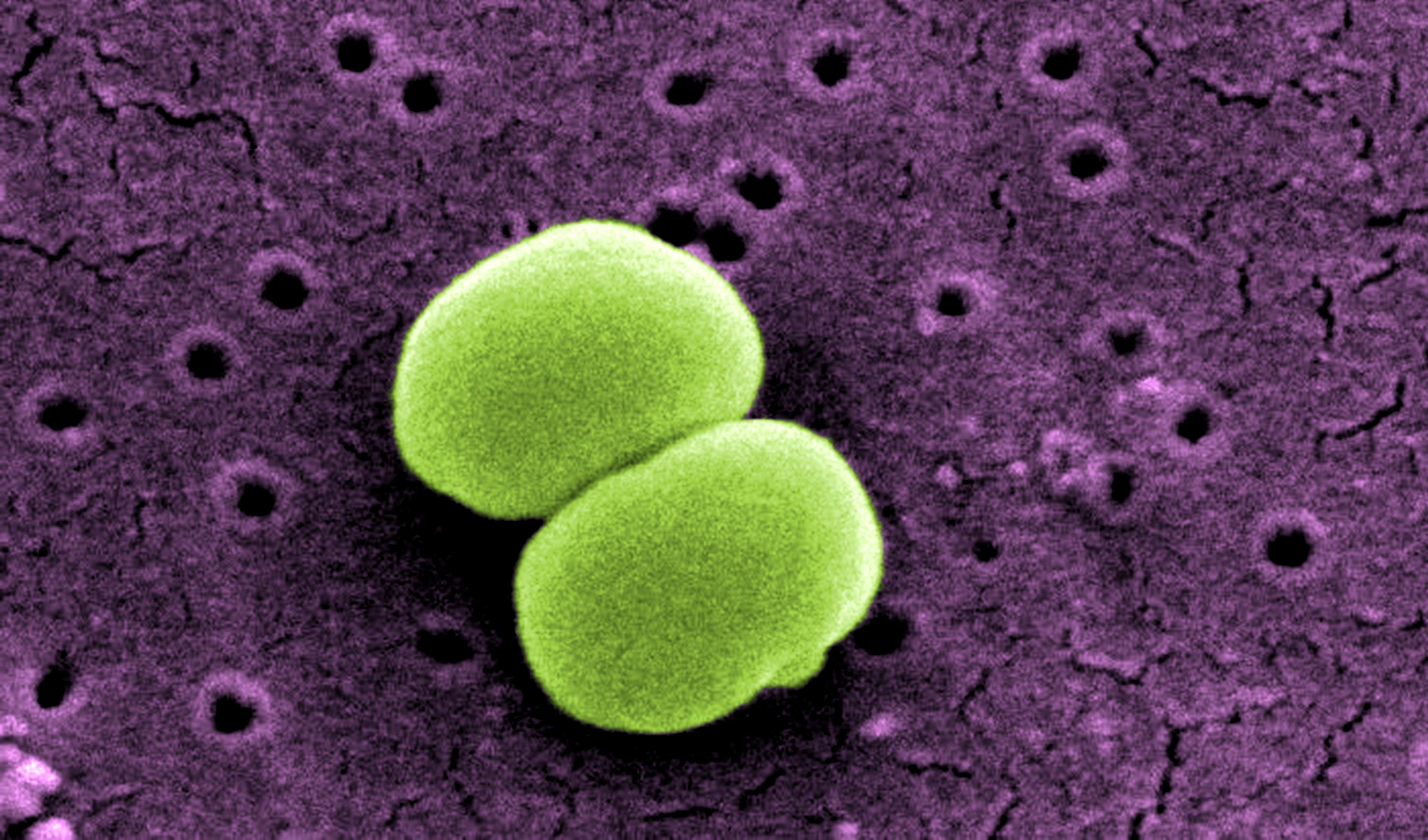
Free picture: scanning, electron micrograph, two ... from pixnio.com 1024 x 768 jpeg 397 кб. Conidiophores are produced by ascomycetes whereas sporangiophores are produced by zygomycetes. 200 x 133 jpeg 16 кб. History and etymology for conidiophore. 282 x 432 jpeg 110 кб. Is the zygospore diploid or haploid? The hyphae were either simple or branched. Be able to identify the conidiospores and conidiphores of the ascomycota specimens.
Conidiophores branching at tips and is darkly pigmented (dark gray when viewed through dissecting microscope).
Vector image penicillium slide ( blue mold, mycelium and conidiophores). Tagide decarvalho (@nerd.candy) в instagram: .microscopic, macro, microscopy, magnified, closeup, under the microscope, micro photography, close up, science, spore, cell, fungi, fungus, biology, microbiology, ascomycetous fungi, ascomycota, penicillium, ascomycete, conidia, conidium, conidiospore, conidiospores, hypha, hyphae. Endospores are formed by a few genera of bacteria, such as bacillus. 1024 x 768 jpeg 397 кб. Observe a slide of penicillium conidiophores under high power. When viewed under the microscope following culture, the following traits were identified: Which of the following would be virtually indistinguishable under the microscope? Direct microscopic examination (with 10% koh) of broken hair strands showed numerous spores inside as well as outside of the hair strand. The spores are called conidia. Tons of macroconidia with rough edges. 200 x 133 jpeg 16 кб. Пин на доске under the microscope.
When viewed under the microscope following culture, the following traits were identified: A single, slender, tubular conidiophore. Is the zygospore diploid or haploid? .microscopic, macro, microscopy, magnified, closeup, under the microscope, micro photography, close up, science, spore, cell, fungi, fungus, biology, microbiology, ascomycetous fungi, ascomycota, penicillium, ascomycete, conidia, conidium, conidiospore, conidiospores, hypha, hyphae. Canis is thicker cell wall and m.
Lignocellulolytic activities of a novel strain of ... from file.scirp.org Tagide decarvalho (@nerd.candy) в instagram: Simultaneously, strands were observed under sem, and there were many round spores in and around the hair strand (fig. .microscopic, macro, microscopy, magnified, closeup, under the microscope, micro photography, close up, science, spore, cell, fungi, fungus, biology, microbiology, ascomycetous fungi, ascomycota, penicillium, ascomycete, conidia, conidium, conidiospore, conidiospores, hypha, hyphae. The hyphae were either simple or branched. First known use of conidiophore. Endospores are formed by a few genera of bacteria, such as bacillus. Which of the following would be virtually indistinguishable under the microscope? Difference between phialides and metulae.
.microscopic, macro, microscopy, magnified, closeup, under the microscope, micro photography, close up, science, spore, cell, fungi, fungus, biology, microbiology, ascomycetous fungi, ascomycota, penicillium, ascomycete, conidia, conidium, conidiospore, conidiospores, hypha, hyphae.
882 отметок «нравится», 15 комментариев — dr. Penicillium, blue mold, mycelium and conidiophores, w.m. Be able to identify the conidiospores and conidiphores of the ascomycota specimens. Post a question or comment about what for example, if you are looking for what stachybotrys chartarum spores and growth structures or conidiophores look like under. Which of the following would be virtually indistinguishable under the microscope? Simultaneously, strands were observed under sem, and there were many round spores in and around the hair strand (fig. Conidiophore and cleistothecium are key factors of penicillium that play a significant role in taxonomic description. Conidiophores are produced by ascomycetes whereas sporangiophores are produced by zygomycetes. 1874, in the meaning defined above. Mold spores under the microscope photo id library of common molds found in buildings. 1024 x 768 jpeg 397 кб. A nucleate, asexual, immotile spore that is generally formed at the apex or side of a specialized sporogenous (conidiogenous) cell. Specialized hyphal strands that produce conidia.
Difference between phialides and metulae conidiospore. Penicillium conidiophores and conidia x 400.